Introduction
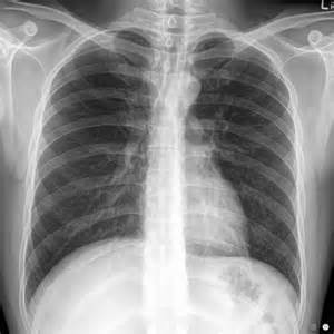
Chest X-ray is performed to visualise organs and structures within the chest cavity such as the lungs, heart, diaphragm, ribs and the main blood vessels.
Routinely, the examination is performed with the patient standing. The patient will be instructed to take in a deep breath and hold it for a few seconds until the X-ray is taken.
 Picture 1: Radiograph (image) of a chest X-ray
Indications
- The doctor will request a chest X-ray if the patient is having the following signs and symptoms:
– Prolonged cough
– Chest pain
– Injury to the chest area
– Coughing of blood
– Difficulty in breathing
- The examination will also be performed on patients with symptoms of tuberculosis (TB), cancer and other lung diseases.
Preparation
- No special preparation is needed for a chest X-ray.
- Necklace and other materials around the chest area need to be removed to avoid them from obscuring the image on the radiograph.
- Patients need to change into a hospital gown that will be provided.
- The examination will be performed by a radiographer.
- Please inform your doctor and the radiographer if you are pregnant.
- Usually the examination will be postponed if you are pregnant or may be pregnant.
| Last Reviewed | : | 5 January 2017 |
| Translator | : | Daud bin Ismail |
| Accreditor | : | Irene Tong Lee Kew |


