

Defintion
Protein electrophoresis is a process that separates proteins based on their size and electrical charge. This forms a characteristic pattern of bands of different widths and intensities on a test media and reflects the mixture of proteins present in the body fluid evaluated. For serum protein electrophoresis, the pattern is divided into five small fractions:
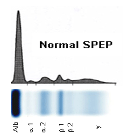
Figure 1 : Protein electrophoresis electrogram patterns
- Albumin (Alb)
- Alpha 1 (?1)
- Alpha 2 (?2)
- Beta, (?) :In some cases, the beta fraction is further divided into beta 1, (?1) and beta 2, (?2).
- gamma ()
Why the test is performed?
Protein electrophoresis is used to identify the presence of abnormal proteins, to identify the absence of normal proteins, and to determine when different groups of proteins are present in unusually high or low amounts in blood or other body fluids.
Immunofixation electrophoresis (IFE) can be used as needed to further identify abnormal bands, in order to determine which type of immunoglobulin is present. Alterations to the usual appearance of the patterns formed can help in the diagnosis of disease.
The presence of an abnormality on a protein electrophoretic pattern is seldom diagnostic in itself. Instead, it provides a clue. In certain circumstances, it can provide as mean of diagnostic for example in multiple myeloma. It can be a supportive evidence for diagnosis in CSF for example, multiple sclerosis. Follow-up testing is then usually performed, based on that clue, to try to identify the nature of the underlying disease. It is usually helpful in monitoring diseases during treatment.
Diagnotic Usefulness
Serum protein electrophoresis is useful to screen diseases for abnormality seen in total protein and/or albumin level, elevated urine protein levels, elevated calcium levels, or low white or red blood cell counts.
The test may be ordered when symptoms suggest a bone pain, anemia, fatigue, unexplained fractures or recurrent infections. The intention is to look for the presence of a characteristic band called paraprotein. If a sharp band is observed, its identity as a monoclonal immunoglobulin is confirmed by immunofixation electrophoresis. Once the disease has been diagnosed, the serum protein electrophoresis test may be ordered at regular interval to monitor the treatment on patient and to see if the monoclonal band is reduced or disappeared completely with treatment. This test is also useful when symptoms suggest an inflammatory condition, an autoimmune disease, an acute or chronic infection, a kidney or liver disorder, or a protein-losing condition.
Urine protein electrophoresis may be used to determine whether the protein is escaping from the blood plasma, suggesting compromised kidney function or is an abnormal protein coming from a different source such as a plasma cell cancer like multiple myeloma. When multiple myeloma is suspected, it is to determine whether any of the monoclonal immunoglobulins or fragments of monoclonal immunoglobulin are escaping into the urine. Its identity is confirmed by immunofixation electrophoresis if a sharp band suggestive of paraprotein is observed. Urine protein electrophoresis also can determine whether the diseases are glomerular or tubular related-diseased in kidney function.
The presences of multiple distinct bands in the CSF, which are present in CSF and not in serum are referred as oligoclonal bands. Other inflammatory conditions of the brain, also present oligoclonal bands. The test can be used to diagnose neurologic symptoms to look for protein suggestive of inflammation, infection or multiple sclerosis.
Additional tests to perform
- albumin
- immunoelectrophoresis
- serum free light chains (Free Kappa and Free Lambda)
- quantitative immunoglobulins
- alpha-1 antitrypsin
- cryoglobulins
Service Available
In Malaysia, 4 hospitals under Ministry of Health and 3 university hospitals offer the Protein Electrophoresis test :
- Hospital Kuala Lumpur (HKL), Kuala Lumpur
- Hospital Ampang, Selangor
- Hospital Queen Elizabeth, Kota Kinabalu, Sabah
- Institute of Medical Researchers (IMR), Kuala Lumpur
- University Malaya Medical Centre (UMMC), Kuala Lumpur
- University Science Malaysia (USM) and
- UKM Medical Centre (UKMMC), Selangor.
Sample Requirement
Samples requirement are different base on the centre that offered the test. The difference is due to the analyzers, methodology and principal applied. However, in order to avoid protein degradation, all the samples need to be kept cold during transportation. Types of sample for protein electrophoresis included
- Blood serum
- Urine
- Cerebral Synovial Fluid (CSF) or
- Synovial fluid
|
Specific Proteins Test
|
Method
|
Normal Values
|
AT (working days)
|
|---|---|---|---|
| Serum Total Protein | Biuret | 58.0 – 82.0 g/L | 2 |
| Urine Total Protein | Pyrogallol red molybdate (PRM) | < 0.2 g/24 hr | 2 |
| Serum Electrophoresis | Agarose-gel electrophoresis (AGE) | Qualitative. | 10 |
| Urine Electrophoresis | Agarose-gel electrophoresis (AGE) | Qualitative | 10 |
| Protein Electrophoresis ( Screening Profiling) – serum – urine |
|
12 | |
| Immunofixation Electrophoresis – serum – urine |
Agarose-gel immunofixation electrophoresis (AGIE) | Qualitative | 20 |
| Paraprotein Quantitation |
|
Absent | |
| Protein Electrophoresis (Confirmatory Profiling) – serum – urine |
|
25 | |
| Total Protein – CSF |
Pyrogallol red molybdate (PRM) | 0.15 – 0.45 g/L | 2 |
| Electrophoresis – CSF Oligoclonal Band |
Immuno-isoelectric focusing electrophoresis | Qualitative | 10 |
Accessed from http://www.imr.gov.my/en on 4th June 2015
Interpretations- The Parameters/ Electrophoregram
The protein electrophoresis test gives a health practitioner a rough estimate of how much of each protein fraction is present and whether any abnormal proteins are present. The value of immunofixation electrophoresis is in the identification of the presence of a particular type of immunoglobulin.
- Serum and urine protein electrophoresis
Albumin is the major protein component of serum and when viewed on the serum protein electrophoresis patterns, it appears to have the largest peak lying closest to the positive electrode. On the other hand, globulin comprises a much smaller fraction of the total content of serum protein. These globulins are the ones which are labeled as alpha 1, alpha2, beta and gamma.Figure 2 exhibits normal and abnormal serum protein electrophoresis and Figure 3 shows the application of serum and urine protein electrophoresis in determination of M component and Bence Jones protein in patient samples.
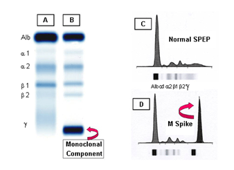
Figure 2 : Normal and abnormal serum protein electrophoresis patterns. A: Normal B: Multiple myeloma showing M component in the gamma region. C: Densitometry tracing of A showing the 5 zones of the high resolution agarose electrophoresis. D: densitometry of the M component of B, termed the M spike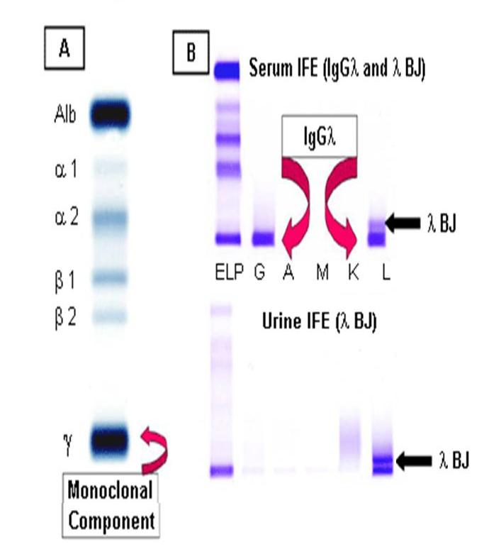
Figure 3 : A:Serum protein electrophoresis demonstrating the M component ; B:Serum and urine immunofixation electrophoresis. Bence Jones protein (? BJ) is present in the urine IFE as well -
Some of the conditions or diseases that may associated with decreases or increases in various serum proteins.
ProteinMay be decreased in:May be increased in:Albumin - Malnutrition and malabsorption
- Pregnancy
- Kidney disease (especially nephrotic syndrome)
- Liver disease
- Inflammatory conditions
- Protein-losing syndromes
- Dehydration
Alpha 1 globulin - Congenital emphysema (alpha-1 antitrypsin deficiency, a rare genetic disease)
- Severe liver disease
- Acute or chronic inflammatory diseases
Alpha 2 globulin - Malnutrition
- Severe liver disease
- Hemolysis
- Kidney disease (nephrotic syndrome)
- Acute or chronic inflammatory disease
Beta globulin - Malnutrition
- Cirrhosis
- High blood cholesterol (hypercholesterolemia)
- Iron deficiency anemia
- Some cases of multiple myeloma or monoclonal gammopathy of unknown significance (MGUS)
Gamma globulin - Variety of genetic immune disorders
- Secondary immune deficiency
- Polyclonal, antibody produced by or derived from different plasma cells:
- Chronic inflammatory disease
- Rheumatoid arthritis
- Lupus
- Cirrhosis
- Chronic liver disease
- Acute and chronic infection
- Recent immunization
- Monoclonal, antibody produced by or derived from a single type (clone) of plasma cell:
- Malignancy
- Multiple myeloma
- Lymphoma
- Waldenstrom’s macroglobulinemia
- ?-cell leukemia
Accessed from https://labtestsonline.org/ on 11 June 2015
- CSF protein electrophoresis
Unlike serum protein electrophoresis which separates proteins base on 5 or 6 fragments, CSF protein electrophoresis is a qualitative test and involve comparison of IgG pattern present in CSF and serum sample. CSF protein is produced by the choroid plexus in the central nervous system. Systemic immunoglobulin cannot transverse blood brain barrier to enter the central nervous system except IgG. Any increased in systemic IgG can be raised in central nervous system because it crosses the blood brain barrier. In multiple sclerosis, there is a productivity of proteins in the central nervous system which is not found in the systemic system. Hence, the protein band can be seen only in CSF not in the serum when analyzing the CSF electrophoresis gel. The migration pattern and electrophoresis profiles are shown in the picture below (Figure 4).
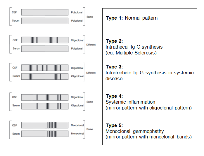
Figure 4 : CSF migration pattern and electrophoresis profiles.
References
- Alper, C., & Johnson, A. (1969). Immunifixation electrophoresis: A technique for the study of paraprotein polymorphism. Vox Sang, 17:445-452.
- Bossuyt, X., Bogaerts, A., Schiettejatte, G., & Blanckaert, N. (1998). Serum protein electrophoresis and immunofixation by a semiautomated electrophoresis system. Clinical Chemistry, 944-949.
- Cawley; L.P; et al.,. (1976). Immunofixation Electrophoretic Technique Applied to Identification of Proteins in Serum and Cerebrospinal Fluid. Clin Chem, 22:1262-1268.
- Lakshminarayanan, R., Li, Y., Janatpour, K., Beckett, L., & Jialal, I. (2007). Detection by Immunofixation of M Proteins in Hypogammaglobulinemic Patients With Normal Serum Protein Electrophoresis Results. American Society for Clinical Pathology, 127:746-751.
- McCudden, C., Mathews, S. P., Hainsworth, S. A., Chapman, J. F., Hammeett-Stabler, C. A., Willis, M. S., & Grenache, D. G. (2008). Performance comparison of cappilary and agarose gel electrophoresis for the identification and characterization of monoclonal immunoglobulin. American Society for Clinical Pathology, 129:451-458.
- O’Connell, T., Horita, T. J., & Kasravi, B. (2005). Understanding and interpretaing serum protein electrophoresis. American Family Physician, 105-112.
- Oda RP et al. . (1997). Capillary electrophoresis as a clinical tool for the analysis of protein in serum and other body fluids. . Electrophoresis, 18, 1715-1723.
- SEBIA. (2012). HYDRAGEL 1, 2, 4 & 9 IF – 2012/03 Product Insert.
- SEBIA. (2012). HYDRAGEL 7, 15 & 30 PROTEIN(E) – 2012/03 Product Insert.
- Waters, J. (n.d.). Correlational Research Guidelines. Retrieved from Capilano University: http://www.capilanou.ca/psychology/student-resources/research-guidelines/Correlational-Research-Guidelines/
| Last Reviewed | : | 23 February 2017 |
| Writer | : | Siti Suhana bt. Abdullah Soheimi |
| Accreditor | : | Chua Peck Ham |


