

What is a MRI BRAIN?
The Magnetic Resonance Imaging (MRI) examination of the brain produces cross – sectional images of the brain that are visualised in the computer. MRI uses magnetic fields and radio frequency waves to produce images of the brain or head. MRI does not have radiation effects as in X-ray.
How and where can I get this examination?
- When you are seeing your doctor who is treating you, the doctor will decide if the MRI examination required.
- If necessary, the doctor will make the request for the examination using the Radiological Examination Request Form.
- This examination is available in selected government hospitals only.
Reasons for performing MRI Brain
MRI brain is performed to diagnose the following conditions;
- Tumour.
- Nerve damage.
- Other abnormalities tin the brain.
Preparation patient prior to MRI Brain examination?
- Normally when you are given an appointment for a MRI Brain examination, you will given the following instruction;
– Fasting 4 – 6 hours before the examination.
– Only plain water can be taken.
– To take allergy medication (steroids) if necessary.
– Inform the doctor who is treating you or radiographer if you have previously experienced the following:
> Asthma.
> Allergy (allergies to food or medications , itching, redness or swelling).
- If you are on any medication, please consult your doctor.
- Please inform the doctor and/ or the radiographer if you are pregnant or suspect that you may be pregnant.
On the day of the examination
Register yourself at the registration counter.
Before examination
- You will be instructed to change into the hospital gown.
- You need to remove any metallic objects including decorative ornaments such as hair pins, watches, dentures, hearing aids, ATM cards, credit cards, pen, spectacles and others. It is feared that these items will be damaged or affect the image quality of the MRI scans.
- Please inform the doctor if you are usig a cochlear implant, metallic joint prosthesis, clips used in brain surgery; aneurysm and cardiac pacemaker as they may be affected by the strong magnetic field.
- Your personal belongings are your responsibility (you are adviced not to bring them along on the day of the examination).
- Please inform your doctor and / or your radiographer if your are or suspect that you may be pregnant.
- Please inform the doctor if you may have any allergy to seafood/ medication or others.
- You will be asked on your preparation, referring to the MRI checklist provided during the appointment.
- Ensure your doctor has discussed with you and filled up the MRI checklist form.
- If you do not conform to the checklist requirement, you will not be allowed to undergo the examination.
- A needle (branula) will be inserted into the vein for examination requiring contrast medium injection.
- Your weight will be taken.
- You will be instructed to to relieve yourself (urinate).
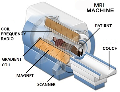
Figure 1: MRI machine
Source: https://www.magnet.fsu.edu
During examination
- The MRI examination will take about 30 – 60 minutes.
- You are instructed to lie on a narrow examination couch, which will later move into position in the bore of the MRI machine.
- If you have claustrophobia (fear of small space), you may require sedatives to calm you down througout the examination.
- During the whole process, you are adviced to calm dowm and remain still throught the whole examination to avoid repeat examination.
- You will be adviced to shut your eye throught the whole examination.
- You will be injected with the contrast medium (if required) at the arm or hand.
- You will hear a knocking sound produced by the MRI machine. There will be an incremental movement of the examination couch into the MRI machine during the scanning process. You are adviced, to remain still in this position.
- The image of the brain/head that is produced is displayed on the monitor.
After examination
- If the examination is long, which requires you to be in the lying down position for, you may experience dizziness when you sit up once the examination is completed.
- You can change into your own clothes. Any needle / branula will be removed.
- You will be allowed to leave after the examination and you are well.
- You are allowed to eat and drink.
Discussion on the follow-up treatment will be done by the doctor with the patient or next of kin.
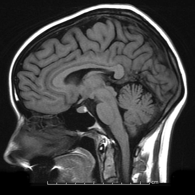
Figure 2: Cross sectional image of the MRI Brain
The report of the examination
All images are reviewed by the radiologist and following and report is prepared.
| Last Review | : | 28 July 2017 |
| Writer | : | Pushpa Thevi Rajendran |
| Accreditor | : | Daud bin Ismail |


