

Introduction
During sexual reproduction process, two gametes (ovum and sperm) fuse and combine their genomes to form the next generation (new offspring – zygote). To avoid inevitable doubling of genetic material in every new generation, genome copy number must be reduced by half before the next round of gametes is formed. This reduction is achieved by an unusual type of cell division — meiosis.
Meiosis occurs in ovaries and testes, where a single cell divides twice to produce four cells containing half the original amount of genetic information. These cells are our sex cells – sperm in males, eggs in females.
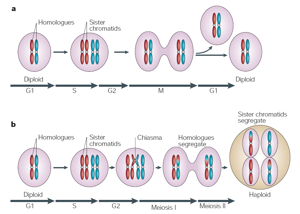
Figure 1: The mitotic and meiotic cell cycles.
© 2004 Nature Publishing Group (Marston, A. L. et al. Meiosis: cell-cycle controls shuffle and deal. Nature Reviews Molecular Cell Biology 5, 984 (2004). All rights reserved.)
Meiosis History
The process of meiosis was first described in the mid-1870s by Oscar Hertwig, who observed it while working with sea urchin eggs. Edouard Van Beneden expanded upon Hertwig’s descriptions, adding his observations about the movements of the individual chromosomes within the germ cells. However, it wasn’t until August Weismann’s work in 1890 that the reduction role that meiosis played was recognized and understood as essential. Some twenty years later, in 1911, Thomas Hunt Morgan examined meiosis in Drosophila, which enabled him to present evidence of the crossing over of the chromosomes.
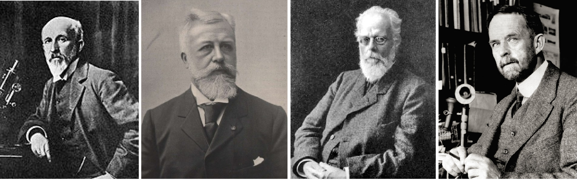
Figure 2: Behind the meiosis discovery: The Scientist
From left: Oscar Hetwig, Edouard Van Beneden, August Weissmann and Thomas Hunt Morgan.
Function
Primary function of meiosis is the reduction of the ploidy (number of chromosomes) of the gametes from diploid (2n, or two sets of 23 chromosomes) to haploid (1n or one set of 23 chromosomes. It promotes genetic diversity and the creation of proper conditions for reproductive success. While parts of meiosis are similar to mitotic processes, the two systems of cellular division produce distinctly different outcomes. Problems occur during meiosis can stop embryonic development and sometimes cause spontaneous miscarriages, genetic errors, and birth defects such as Down syndrome.
Stages of Meiosis
Meiosis can be divided into nine stages. These are divided between the first time the cell divides (meiosis I) and the second time it divides (meiosis II):
Meiosis I
- Interphase:
- The DNA in the cell is copied resulting in two identical full sets of chromosomes.
- Prophase I:
- The copied chromosomes condense into X-shaped structures. At this stage, chromosome can be seen under a microscope.
- Each chromosome composed of two sister chromatids, containing identical genetic information.
- The chromosomes start pairing up together with its own partner, chromosome 1 and chromosome 1, chromosome 2 and chromosome 2, and so on.
- The paired chromosomes may then exchange bits of DNA in a process called recombination or crossing over.
- At the end of Prophase I, the membrane around the nucleus in the cell dissolves away, releasing the chromosomes.
- The meiotic spindle, consisting of microtubules and other proteins, extends across the cell between the centrioles.
- Metaphase I:
- The chromosome pairs line up next to each other along the centre (equator) of the cell.
- Meiotic spindles start extending from centrioles that are at the opposite poles of the cell.
- The meiotic spindle fibers attach to one chromosome of each pair.
- Anaphase I:
- Meiotic spindles pull apart the chromosome pairs where each one chromosome will go to one pole of the cell and the other chromosome to the other opposite pole
- In meiosis I the sister chromatids stay together. This is different to what happens in mitosis and meiosis II.
- Telophase I and cytokinesis:
- The chromosomes complete their move to the opposite poles of the cell.
- Each pole of the cell has a full set of chromosomes gather together.
- A membrane forms around each set of chromosomes to create two new nuclei.
- The single cell then pinches in the middle to form two separate daughter cells each containing a full set of chromosomes within a nucleus. This process is known as cytokinesis.
Meiosis II
- Prophase II:
- Now there are two daughter cells, each with 23 chromosomes (23 pairs of chromatids).
- In each of the two daughter cells, the chromosomes condense again into visible X-shaped structures that can be easily seen under a microscope.
- The membrane around the nucleus in each daughter cell dissolves away releasing the chromosomes.
- The centrioles duplicate.
- The meiotic spindle forms again.
- Metaphase II:
- The chromosomes (pair of sister chromatids) in each of the two daughter cells start to line up end-to-end along the equator of the cell.
- The centrioles are now at opposites poles in each of the daughter cells.
- Meiotic spindle fibers at each pole of the cell attach to each of the sister chromatids.
- Anaphase II:
- The meiotic spindle pulled the sister chromatids to the opposite poles.
- The separated chromatids are now become individual chromosomes.
- Telophase II and cytokinesis:
- The chromosomes complete their move to the opposite poles of the cell.
- A full set of chromosomes gather together at each pole of the cell.
- A membrane forms around each set of chromosomes to create two new cell nuclei.
- This is the last phase of meiosis; however cell division is not complete without another round of cytokinesis.
- Once cytokinesis is complete there are four granddaughter cells, each with half a set of chromosomes (haploid):
- In males, these four cells are all sperm cells
- In females, one of the cells is an egg cell while the other three are polar bodies (small cells that do not develop into eggs).
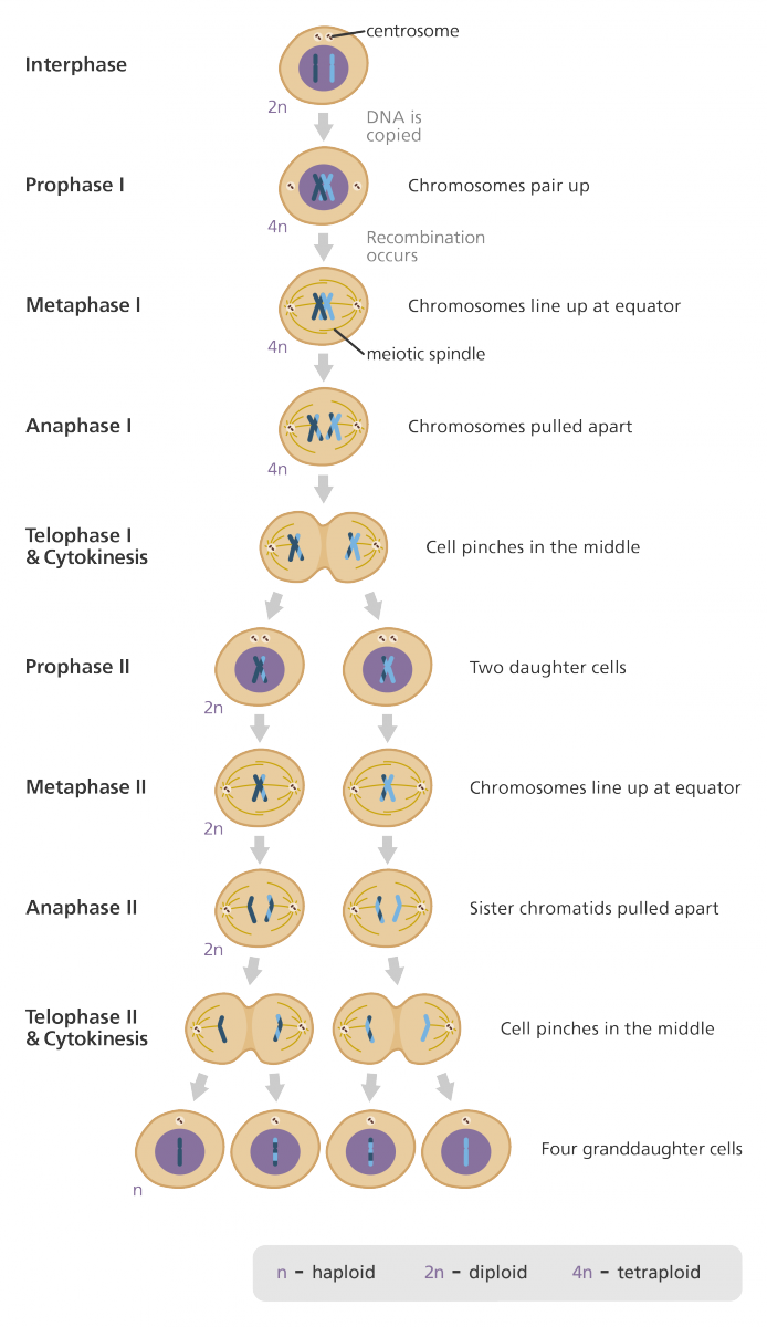
Figure 3: Stages of meiosis.
Image credit: Genome Research Limited
References
- http://www.cell.com/current-biology/fulltext/S0960-9822(08)00733-1
- Marston, A. L. et al. Meiosis: cell-cycle controls shuffle and deal. Nature Reviews Molecular Cell Biology 5, 984 (2004).
- Maayan, Inbar, “Meiosis in Humans”. Embryo Project Encyclopedia (2011-03-24). ISSN: 1940-5030 http://embryo.asu.edu/handle/10776/2084.
- http://www.yourgenome.org/facts/what-is-meiosis
| Last Reviewed | : | 07 April 2017 |
| Writer | : | Siti Zaharah Farah Dura Abu Bakar |
| Accreditor | : | Farah Amalina bt. Mohd Zulkufli |


