

Introduction
Karyology is the study of whole sets chromosome by referring to its standard format known as karyogram or ideogram. Cells collected from individual are induced to divide and the arrested at metaphase stage (a stage in cell division) where the chromosomes are condensed and visible. Human somatic cells comprise of 23 pairs of chromosome (22 pairs autosome and one pair sex chromosome). This makes a total number of 46 chromosomes. The different between male and female is the inheritance of the sex chromosome pair; either XX or XY. Therefore normal female karyotype will have 46,XX (Figure 1(a)) whereas male is 46,XY (Figure 1(b)).
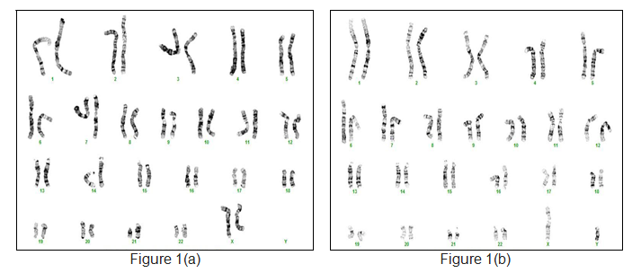
Figure 1(a) showed normal female karyotype of 46,XX whereas Figure 1(b) represent normal male 46,XY
The chromosomes are stained with certain dyes resulting in light and dark bands called banding pattern. These banding patterns and chromosome sizes are used in chromosomes identification.
History of Karyotype Analysis
Chromosomes were first observed in 1842 by Carl Wilhelm von Nägeli which later being further described in salamander by Walther Flemming and Heinrich von Waldeyer. Development of this field took another step in early 20th century when chromosome was started to be pointed as the carrier of the genes that causes the phenotypic (physical) characteristics of the cells.
After many years of research, in 1912 Hans von Winiwarter reported 47 chromosomes in spermatogonia and 48 in oogonia thus concluding an XX/XO sex determination mechanism. Later in 1956, Joe Hin Tjio and Levan proposed that complete human karyotype only composed of 46 chromosomes in somatic cells. He also proposed several approaches in karyotype analysis such as;
- Using cells in tissue
- Pretreatment of cells in hypotonic solution
- Usage of colchicines to arrest mitosis in metaphase stage
- Slide preparation by squashing chromosomes onto a single plane
- Cutting up photomicrograph and chromosome classification according to respected karyogram
Purpose of Karyotype Analysis in Human
Karyotype analysis is commonly used for chromosomal identification (karyotyping) and also to detect abnormalities involving chromosome number as well as changes in chromosome structure (translocation, deletion, addition etc.). Detection of chromosomal changes through karyotyping often associated with syndromes such as trisomy 21 or Down Syndrome where individual having 3 copies of chromosome 21 (extra one copy). Changes in chromosome structure also provide information about changes of genes that may lead to certain diseases. For example, translocation between chromosome 9 and 22 causes leukaemia cancer cells, particularly chronic myelogenous leukaemia (CML). Understanding genetic material changes facilitates diagnosis, prognosis and patient management.
Samples Used
Obtaining chromosome is the main goal in this karyotype analysis and the entire parts of human body contains this source of DNA. Samples can be collected from:
- Blood
- Saliva
- Tissue
- Teeth
- Bones etc.
Techniques in Karyotype Analysis
To begin karyotype analysis, source of DNA (i.e. peripheral blood) is collected from individuals, followed by initiation of cell culture in growth medium for several days. Chromosomes are obtained by several steps of harvesting which includes hypotonic solution treatment and fixations. Chromosomal suspension is dropped onto slides and banded with specific staining method resulting in appearance of banding pattern on each chromosome. Metaphase spreads is then visualized, captured and analyze using software analyzer (Figure 2).

Figure 2 illustrate a G-banded metaphase spread viewed under light miscroscope
Banding Technique
After the first step by Tjio and Levan reporting diploid number of human chromosome, several banding techniques have been proposed which led to a standard nomenclature in classification of human chromosome. These banding techniques are used depending on the purpose as well as the targeted region to be observed on chromosome:
- Giemsa banding (G-banding)
- Reverse banding (R-banding)
- Centromere banding (C-banding)
- Quinacrine banding (Q-banding)
- Telomere banding (T-banding)
- Nucleolar organizer region banding (NOR-banding)
Giemsa banding and reverse banding are widely used techniques that produces banding pattern composing of dark and light transverse bands on the chromosome. In Giemsa banding (G-banding), dark region represent heterochromatic area with less replication than lighter region of euchromatic region (Figure 3(a)). In contrast with G-banding, dark band in reverse banding (R-banding) is euchromatic region while light region is heterochromatic region (Figure 3(b)). Banding pattern allows homologous identification and construction of chromosomal nomenclature for many species. Comparison between chromosome pair and standard karyogram is used to identify any segmental changes.
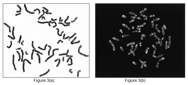
Figure 3(a) showed G-banded metaphase spread while Figure 3(b) showed R-banded metaphase spread
Observation on Karyotypes
Chromosome analysis involves observation, evaluation and comparison of copy number, structure and banding pattern of each chromosome with the standard ideogram or karyogram. In human karyotyping, several characteristic is observed involving (Figure 4);
- Relative size of chromosome
- Position of centromeres
- Basic number of chromosomes
- Number and position of satellites on acrocentric chromosome
- Banding pattern related to distribution of heterochromatic and euchromatic region
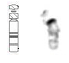
Figure 4 showed normal chromosome 22 (right) that was arranged next to its standard ideogram (left)
Limitations of Karyotype Analysis
Karyotype analysis detects nucleotide changes of 5 Megabases (5 x 10⁶ bases) and above. For example, Down Syndrome patients have a total number of 47 chromosome due to the presence of extra copy of chromosome 21 (Figure 5(b)). The same goes with classical Turner Syndrome where the there is loss of one chromosome X making the total number of chromosome in the cell is 45 rather than 46 chromosomes in normal cells (Figure 5(c))
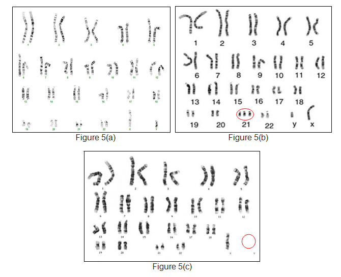
Figure 5(a) showed normal male karyotype of 46,XY; sporadic Down Syndrome in Figure 5(b) having extra copy of chromosome 21 and loss of another chromosome X in proband Figure 5(c) (Turner Syndrome)
Karyotyping relies on G-banding quality and chromosomal resolution. Small structural rearrangements i.e. nucleotide changes of less than 5 Megabases (<5 Mb) in chromosome can be missed by this analysis. Thus, most of geneticists and genetics laboratory will carry other further testing simultaneously to complete this chromosomal investigation.
References
- https://en.wikipedia.org/wiki/Cytogenetics
- http://www.innovateus.net/health/what-karyotype
- http://www.labs.gosh.nhs.uk/media/384333/Limitations%20of%20cytogenetic%20testing.pdf
| Last Reviewed | : | 07 April 2017 |
| Writer | : | Norashikin bt. Abd Muin |
| Accreditor | : | Farah Amalina bt. Mohd Zulkufli |


