

Introduction
Magnetic Resonance Spectroscopy (MRS) is a non-invasive diagnostic test of measuring biochemical changes from the brain, especially the presence of tumors. While magnetic resonance imaging (MRI) identifies the anatomical location of a tumor, MRS compares the chemical composition of normal brain tissue with abnormal tumor tissue. Tissue changes into stroke and epilepsy also can be detect by using this test.
How does MRS work?
The basic principle that enables MRS is the electron cloud of an atom that shields the nucleus of the magnetic field to a greater lesser degree. This naturally results in slightly resonant frequencies, which in turn return a slightly different signal.
MRS is conducted on the same machine as conventional MRI. The MRI scan uses a powerful magnet, radio waves, and a computer to create a detailed images. Spectroscopy is a series of test that are added to the MRI scan of your brain or spine to measure the chemical metabolism of a suspected tumor.
MRS analyzes molecules such as hydrogen ions and protons. Proton
spectroscopy is more commonly used. There are several metabolites,or products of metabolism, that can be measured to differentiate between tumor types such as:-
- Amino acids
- Lipid
- Lactate
- Alanine
- N-acetyl aspartate
- Choline
- Creatine
- Myoinositol
The frequency of these metabolites is measured in units called parts
per million (ppm) and plotted on a graph as peak of varying height. By measuring each metabolites ppm and comparing it to normal brain tissues, the neuroradiologist can determine the type of tissue present. The example peaks of tumor types are:-
- Lactate peaks : resonates with 1.3 ppm
- Lipid peaks : resonates with 1.3 ppm
- Alanine peaks : resonates with 1.48 ppm
- N-acetyl aspartates : resonates with 2.0 ppm
- Choline peaks : resonates with 3.2 ppm
- Creatine peaks : resonates with 3.0 ppm
- Myoinositol peaks : resonates with 3.5 ppm
What does a MRS show?
MRS spectroscopy can be used to determine tumor type and Aggressiveness, and distinguish between tumor reccurrence and Radiation necrosis. Different metabolites can indicate:-
- Glioma : lower than N-acetyl aspartate levels, elevated
choline and lipid levels, and lactate peaks. - Radiation necrosis : does not have elevated choline level
- Meningioma : elevated alanine level
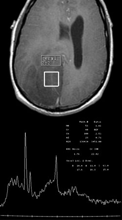
Figure 1 : MRS graph shows the different chemical peaks of a suspected brain tumor
What are the risk?
MRI and MRS are very safe. There are no known health risk associated with the magnetic field or the radio waves used by the machine. Some people are sensitive to the contrast agent and may develop an allergic Reaction. All contrast agents are safe.
Some special circumstances limit the use of a magnetic field. So, it is Important to tell your doctor if any of the list applies to you:-
- Cardiac pacemaker
- Metal plate, pin, or metallic implant
- Intrauterine device
- Insulin or other drug pump
- Cochlear implant
Any metallic substance on your body can affect the quality of the images and values obtained. It can also cause discomfort or injury to you when placed in the magnetic field, and may exclude you from the exam.
You should also inform your healthcare team if you are pregnant. The American College of Radiology does not recommend MRI scanning during the first trimester of pregnancy. You must need consent to the specialist to proceed with this test to be performed.
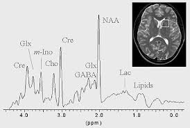
Figure 2 : Investigation of brain injury by MRI and MRS
References
- Majos, C. et al. Proton MR Spectroscopy Improves Discrimination between Tumor and Pseudotumoral Lesion in Solid Brain Masses. American Journal of Neuroradiology 30:544-551, 2009
- Sanghvi, D. Recent advances in imaging of brain tumors. Indian Journal of Cancer 46:82-87, 2009
- Nelson SJ, McKnight TR, Henry RG. Characterization of untreated gliomas by magnetic resonance spectroscopic imaging. Neuroimag Clin 12:599-613, 2002.
- http://appliedradiology.com/articles/advanced-mr-techniques-in-brain-tumor-imaging
| Last Reviewed | : | 17 March 2017 |
| Writer | : | Nik Mohd Amiruddin bin Nik Pakheruddin |
| Accreditor | : | Adzlin Hana bt. Mohd Sari |


