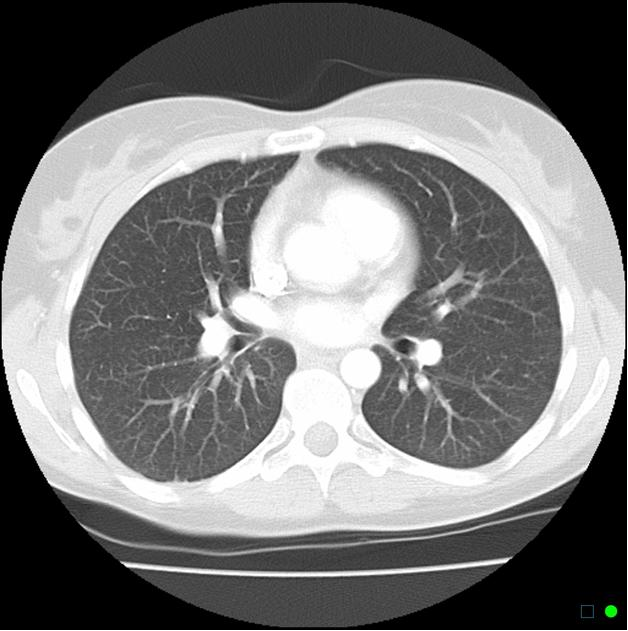
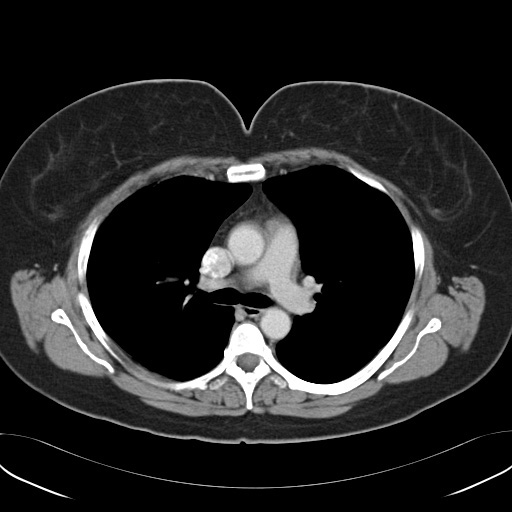
What is CT Thorax?
Computed Tomography (CT) Thorax is a scan to visualise the morphological structures of the organs in the thoracic cavity such as the heart, major blood vessels, lungs, pleural cavity and other organs in the mediastinum and upper part of abdominal cavity such as the liver.
Reasons for a CT Thorax
The CT Thorax examination is performed to diagnose;
- Accumulation of blood or fluid in the lungs
- Infection of the lungs
- Tumour
- Other abnormalities that increases the fluid content in the lungs when contrast medium is injected.
How and where can I Get this examination?
- When you visit the doctor treating you, the doctor will decide if the examination is required.
- If required, the doctor will request for the examination using the Radiology Examination Request Form.
- This examination is available in selected MoH hospitals.
Before a CT Thorax examination
- The radiographer will explain the procedure to you
- Inform your doctor or radiographer if you are pregnant or suspect that you may be pregnant
- If the examination requires the use of contrast medium, there a few instructions that need to be adhered such as the following:
– You have to fast a minimum of 4 – 6 hours before the procedure.
– Take the steroid medication that is supplied 12 hours and 2 hours before the examination.
– You are adviced not to wear or bring along any valueables on the day of the examination.
- You have to remove all metalic objects such as hair clips, earings, chain and others to avoid artifacts on the image.
- If your CT Thorax examination requires contrast media, then your doctor will insert a needle (branula) into your vein at the hand or arm.
- Clothes or accessories with metallic object in the thoracic area (eg: chain, bra and others) have to be removed and you will be need to change into the hospital gown.
- Inform your doctor or radiographer if you are pregnant or suspect that you may be pregnant.
- Your weight will be taken.
During examination
- You will be instructed to lie on the examination couch.
- The radiographer will give clear intruction of the examination that need to be followed.
- The radiographer will position your body and you need to keep still in the position.
- The examination couch will be moved into the gantry.
- The radiographer will be in the control room to operate the CT scanner.
- The CT scanning will start.
- Although the examination room door is closed, you are still able to communicate with the radiographer/ or doctor via intercom.
- You are adviced to not move throughout the procedure (CT Thorax) to avoid distortion to the image which will cause examination to be repeated.
- If contrast medium is required during the examination, you may experience discomfort when the contrast medium is injected into the vein such as sudden warm sensation, nausea, dizziness and others. However all these symptoms are temporary.
- The doctor will be continuously monitoring your status to ensure you are fine.
- The images produced will be displayed on the monitor.
- On completion of the examination, the examination couch will be moved out of the gantry.
- You are allowed to get off the examination couch and change into your clothes.
After examination
- You can change into your clothes.
- If there was a needle(branula) inserted by the doctor, ensure that it is removed before leaving.
- On completion of the examination, please adhere to the apponiment date given by the requesting doctor.
- For those who have has the plain CT Thorax (without contrast medium) there is no specific post procedure care.
- For those who has CT Thorax with contrast medium injection into the vein you are adviced to drink lots of water to speed up the process of excretion of contrast medium from your body. Through urine.
- Ensure that you are stable before you leave the department.
- If you do experience any problems, please do inform the radiographer on duty.
Examination report
All images produced are reviewed by the radiologist and report is prepared.
Figure 1 (a), (b): Normal CT thorax image.
| Last Review | : | 27 July 2017 |
| Writer | : | Pushpa Thevi Rajendran |
| Accreditor | : | Daud bin Ismail |


