Overview
Root canal treatment is performed to save teeth with injured pulp from extraction, so that it won’t be extracted and can remain functional in the mouth. In some cases, where the reinfection occurs or persists in root treated teeth, root canal retreatment is needed. Normally, this non-surgical procedure will be sufficient to heal the tooth. Occasionally due to several factors, retreatment is done followed by periapical microsurgery. Sometimes this surgery is recommended in cases where non-surgical root canal retreatment is unable to be carried out due to some reasons such as in cases with post crown.
What is a periapical microsurgery?
Periapical is referred to area surrounding the tip of the root. Periapical microsugey is also known as endodontic microsurgery or apicoectomy. It is a minor surgery where a small incision is normally made in the gum tissue related to involved tooth in order to gain access to infected area and to expose the bone and surrounding inflamed tissue. All these procedures are performed after profound local anaesthetic is achieved. The infected or inflamed tissue is removed and root tip is prepared or resected when necessary. A root-end filling is then placed at the resected and prepared part of the root tip to prevent re-infection of the root before the gum is sutured back in place. The bone naturally heals around the root over a period of two months restoring its full function.
Several X-rays of the tooth and surrounding bone will be taken before and after the surgery. This procedure is usually carried out under special microscope and ultrasonic instruments to provide better visibility and accessibility respectively. This increases the chance that the procedure will succeed.
Following the procedure, there may be some discomfort or slight swelling while the incision heals. This is normal for any surgical procedure. To alleviate any discomfort, an appropriate medication will be recommended.
Why Periapical Microsurgery Is needed?
Periapical microsurgery is a promising procedure if a previously root canal treated tooth exhibits signs or symptoms of persistent or recurrent infection. It is indicated in cases with periapical pathosis that are related to a periapical cyst, a complex canal anatomy, extraradicular infection, an inadequate healing after nonsurgical retreatment.This microsurgery can also be used to locate root fractures or hidden canals that do not appear on x-rays but still manifest pain in the tooth especially if it has a large post and satisfactory crown coverage. In addition, it may be needed to remove separated instrument within root canal, and to treat damaged root surfaces or the surrounding bone
What are the possible risks and complications?
Like other surgery, there are possibilities of risks and complications in performing periapical microsurgery. The procedures involving posterior teeth (premolar and molar) are more challenging compared to those in anterior region Roots of the upper posterior teeth are often in close proximity to the floor of the maxillary sinus which can be perforated during the procedure. Paraesthesia or prolonged numbness in the mandibular arch might occur after periapical microsurgery of the lower teeth. The reason is due to close relation of the roots to the mental foramen where the mandibular nerve is passing through. It is also difficult to get a good apical seal in posterior teeth because of their root anatomy and position..
Procedures In Periapical Microsurgery
 |
|
 |
|
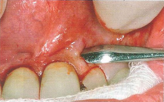
1. Elevation of the gum after incision is made1
|
|
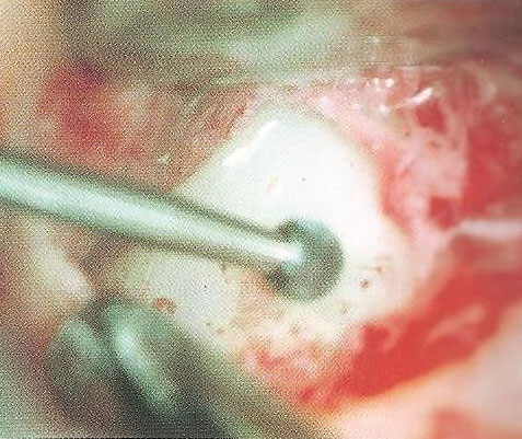
2. Removal of bone to expose the apical area1
|
|
|
|
|
 |
|
 |
|
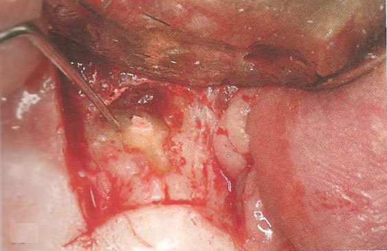
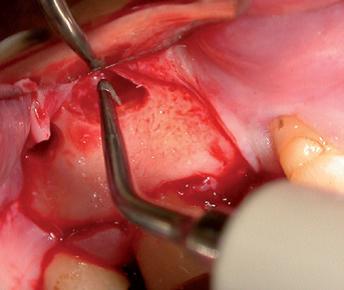
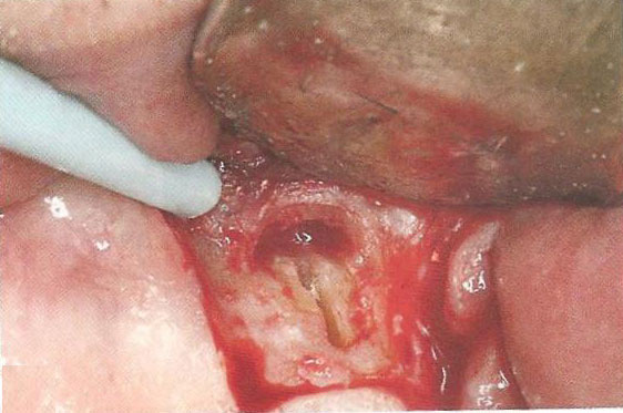
3. Exposure of root apex and the lateral surface of root1
|
|
4. Use of specially designed ultrasonic tip to prepare root end cavity2
|
|
|
|
|
 |
|
 |
|
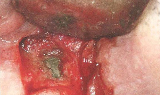
5. Prepared apical canal space1
|
|
6. Root end filling (Mineral Trioxide Aggregate) in prepared apical canal space1
|
References
- James L. Gutmann & Paul E. Lovdahl. Buku Teks: Problem Solving In Endodontics: Prevention, Identification And Management 5th Edition: 325-355
- B. S. Chong and J. S. Rhodes. Endodontic surgery. Br Dent J 2014 Mar 21; 216 (6): 281-290
| Last Reviewed | : | 23 August 2019 |
| Writer | : | Dr. Zaini bt. Aziz |
| Accreditor | : | Dr. Chua Meei Jinn |
| Reviewer | : | Dr. Roshima bt Mohd Sharif |


