

Cancer
Cancer is one of the leading causes of death worldwide which contributes to 8.2 million deaths in 2012. There are several types of cancer such as lung, liver, stomach, colorectal and breast cancers that cause the most cancer deaths each year.
 Figure 1: Cancer can cause stress to the patient.
Source of image:
http://www.health.com/health/stress-management/
 Figure 2: Graph types of cancer that occur with age in males and females.Source of image:
http://globocan.iarc.fr/factsheets/populations/factsheet.asp?uno=900
About 30 % of cancer deaths are due to the unhealthy lifestyle and risky eating habit such as overweight or obesity, poor diet, lack of exercise, smoking and excessive alcohol intake. It is expected that annual cancer cases will rise to 13.1 million in 2030.
 http://www.fabuloussavings.com/blog/wp-content/uploads/2010/12/Lazy-Exercise.jpg
Cancer results from uncontrolled cell growth. Cancer becomes dangerous when the cells disrupt the normal function of the body and it is shown by certain symptoms. Usually, these symptoms only appear when the cancer is already severe.
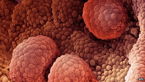 Image 2: Cells from prostate cancer.Source of image http://www.bbc.co.uk/news/health-21628911
There are two categories of cancer which is non cancerous ( benign) and cancerous (malignant ). Cancer cells are more dangerous because it can spread from where it started to other organs or parts of the body systemically ( via the bloodstream or lymphatic system ) and can disable the function of other organs, resulting in death.
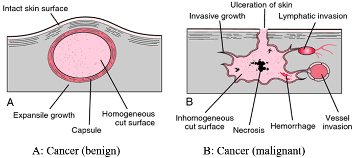 Source of image: http://medical-dictionary.thefreedictionary.com/benign+tumor
Tumour Marker
Tumour marker is a substance that can be measured in the blood, body tissue or other body fluids. Measured values ??can help in the process of detecting and treating cancer patients. It may take the form of proteins, antigens or hormones and others.
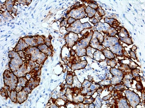 Detection of tumour markers in body tissue histology test .
Source : http://www.epitomics.com/diagnostics/product/5613
- The role of the tumour marker
It is very helpful in determining the type and condition of the cancer (aggressiveness, growth rate, and the stage of cancer). The results of tumour marker test is more useful for cancer diagnosis and treatment of disease when used along with other tests such as radiology tests ( X –ray) or biopsy tissue.
In general, the same type of tumour markers may be present in certain types of cancer that originates from the same tissue. Tumour markers may be elevated in cancer patients, yet sometimes the increase can also occur in patients with tumors ( non-cancerous ) or in the body of a normal person . It is produced by the cancer cells themselves or other cells in the body in response to the presence of cancer.
- The ideal tumour marker
The ideal tumour marker is able to determine the cancer specifically and highly sensitive for the detection of early cancer screening or diagnosis of cancer. However, most tumour markers do not have these features.
Although not very specific or sensitive to certain type of cancer, the high level of tumour marker will be lower after treatment or surgery. Thus measuring tumour marker is helpful to access the effectiveness of treatment or therapy. It is also useful in detecting recurrence of the cancer.
- The need for tumour marker tests.
Tumour marker tests should only be carried out if there is an indication or with medical device. Slight elevation of level will cause unnecessary anxiety or distress to the individual. It will also end up doing unnecessary investigation like colonoscopy, CT scan or MRI which are costly.
 Source: www.medindia.net
Types of Tumour Marker
Today, there are a variety of tumour markers that have been discovered and used in the medical field.
Commonly used tumour marker today is as follows:
• CEA (Carcinoembriogenic antigen)
Carcinoembriogenic antigen ( CEA ) is a protein that may be present in the blood of most patients with cancer, particularly cancer of the large intestine (colon and rectum ). It may also be present due to other cancers such as cancers of the pancreas, breast, ovary, or lung.
Other conditions ( non-cancerous ) that can increase the CEA in the blood which is cirrhosis , hepatitis , inflammatory bowel disease , peptic ulcer disease , or inflammation of the gallbladder ( cholecystitis ) .
In addition, the increase in the levels can also occur in cigarette smokers though no presence of cancer.
CEA is helpful in the detection of recurrent cancer incidence after a patient has undergone treatment. But it is not suitable for screening or diagnosis of cancer is still in its infancy.
• CA 125 ( Cancer Antigen 125 )
CA125 levels will rise when ovarian cancer cells release into the bloodstream of the patient. It is a protein produced by a lot of the surface layer of ovarian cancer cells compared to cells of the body.
Normally CA125 test done to see the effectiveness of the treatment carried out or detect cancer recurrence.
In addition, it is also used to determine the origin of cancer initiation when a woman is diagnosed with cancer in other organs (metastatic cancer) as well as the ovaries.
CA125 is not suitable for use in the screening of cancer. CA125 test results are used in conjunction with the results of an ultrasound scanner can help detect ovarian cancer in women who have a high risk of getting the disease.
• CA 19-9 ( Cancer antigen 19-9 )
Tumour marker CA 19-9 is often used in the treatment of pancreatic cancer patients .
Although it is not very sensitive for the detection of cancer in its early stages, but it is still the best tumour markers for pancreatic cancer patients.
If the patient is newly diagnosed with pancreatic cancer have high levels of CA 19-9 compared to the normal range, it showed the cancer was in stage 3 or 4.
CA19 – 9 is also used for bladder cancer, especially to see the aggressiveness of the cancer. Besides the increase can also be seen in cancers of the digestive tract , particularly the stomach and gallbladder cancer. Non-cancerous conditions that would increase the value of CA19- 9 in a person’s body is thyroid disease, rheumatoid arthritis, inflammatory bowel system or pancreas.
- CA 15-3 ( Cancer antigen 15-3 )
Tumour marker CA 15-3 is widely used for breast cancer. Only 10% of the increase can be detected at an early stage of breast cancer, while it can be detected in 70% of breast cancer patients who are already level 3 or 4.
Levels should be decreased if given effective treatment, but it will increase the first few weeks of treatment. (Excessive happens because the cells that die during the treatment process secrete their contents into the bloodstream).
CA 15-3 can also be detected in other cancers such as cancers of the lung, colon , pancreatic , ovarian and breast cancer ( benign) , ovarian disease , endometriosis and hepatitis .
High levels can sometimes also occur in women who do not have cancer.
• PSA ( Prostate Specific Antigen)
Normal prostate cells produce a protein called Prostate Specific Antigen ( PSA ) . The increase level of PSA than the normal range, indicating the presence of prostate cancer. But it also increased in the presence of harmless tumors of benign prostatic hyperplasia ( BPH ) in addition to acute bacterial infection in the prostate glands or taking certain medications .
PSA test has been widely used in prostate cancer screening program. However, many studies are still underway to determine the effectiveness of its role in cancer screening process.
PSA levels may also reflect the volume of the prostate glands in addition it will also increase with increasing age.
Digital rectal examination (Digital rectums Examination) with PSA tests can help doctors make a diagnosis of prostate cancer more accurately.
• AFP (Alpha – fetoprotein )
Alpha- fetoprotein ( AFP ) is a protein produced during the first three months of fetal development . Then it will drop dramatically when a baby is 1 year old and the levels are very low in the body of a healthy adult.
AFP level are usually higher than normal if someone is suffering from liver cancer or hepatocellular carcinoma. Its value will increase due to germ cell tumour, the disease is rare. Other cancers that would also increase the level of AFP, including Hodgkin’s disease, lymphoma and kidney cancer .
Besides cancer, other conditions that will cause the AFP in the blood are cirrhosis, chronic hepatitis and pregnant women.
• HCG ( human chorionic Gonadotrophin )
HCG is a hormone glycoprotein containing beta subunit ( ? – HCG ) . This hormone increases normally in pregnant women. The increase may be linked to cancer originating from the ovum or sperm cells , such as cancer of the ovary or testis ( germ cell tumor ) and gestational trophoblastic disease , especially Choriocarcinoma.
Under normal circumstances the increase of HCG in women may be associated with pregnancy. It is also very useful in detecting and monitoring ectopic pregnancy, molar pregnancy and miscarriage problems. This hormone will not exist in the body of a healthy man.
Samples and preparation of patients for tumour marker tests
Markers of cancer can be detected by means of qualitative or quantitative blood samples, urine, body tissues or other body fluid .
Patients do not need to fast, blood can be taken from the patient at any time . However, factors such as the period before treatment , medications , age, gender etc. can influence the outcome of tumour marker tests .
 http://melanomablog.co.uk/update-on-vitamin-d/2011/01/30/
Tumour marker test methods
There are several methods used in measuring the level of tumour markers in patient specimens . Among them immunoassay methods, proteomic , genomic or bioinformatics .
 http://www.riqas.com/immunoassay-speciality-I-eqa.php
The selection method is based on the type of tumour markers to be detected and involved either a blood sample , urine , body tissues or other body fluids .
Table 1 below is a summary of the markers that have been described previously. There are also the normal ranges for the tumour markers. However it should be noted that the normal range is only as a guide because it is different between laboratories depending on the methods and analyser used. Your physician is the most appropriate in interpreting the tumour markers measured.
| Tumour Markers | Place primary tumour marker synthesis by cancer cells | false positives | Normal Range |
| Alpha-fetoprotein (AFP) | Liver , germ cells of the ovary or testis | pregnant | 0-6.4 IU / ml
( except pregnant women ) |
| CA15-3 (carbohydrate antigen 15-3) | Breast | < 31 U/ml | |
| CA19-9 | Pancreas , colon –rectal | < 33 U/ml | |
| CA125 | Ovarian | Pregnant, Periods | 0-35 U/ml |
| Carcinoembryonic antigen (CEA) | Colon | Smoking | < 3 ng/ml
< 5 ng/ml (smoker) |
| hCG (human chorionic gonadotropin) | Trophoblastic Disease , Choriocarcinoma | pregnant , smoking, testicular failure | > 31 IU/ml |
| Prostate specific antigen (PSA) | Prostate | < 4 ng/ml |
Table 1
There are a variety of other tumour markers such as anaplastic lymphoma kinase ( ALK ), BCR- ABL, Beta- 2 – microglobulin (B2M) , Bladder tumor antigen (BTA) , BRAF , CA 27-29 , Calcitonin , Chromogranin A, Epidermal growth factor receptor (EGFR).
But some of these markers are not been widely used due to factors such as high cost of testing and still require more research to determine the appropriateness as tumour markers.
References:
- http://www.uihealthcare.org/2column.aspx?id=23746
- Tumor Markes: Physiology, Pathobiology, Technology, Clinical Apllications
Eleftherios P. Diamandis et al. 2002
- http://books.google.com.my/books?id=qwJHDfXMozUC&printsec=frontcover&dq=tumor+marker&hl=en&sa=X&ei=YFHCUby8BonYrQfrqYDoAQ&ved=0CDgQuwUwAA#v=onepage&q=tumor%20marker&f=true
- Pagana KD, Pagana TJ (2010).Mosby’s Manual of Diagnostic and Laboratory Tests, 4th ed. St. Louis: Mosby Elsevier.
- http://www.webmd.com
- http://emedicine.medscape.com/article/457394-overview#a1
- http://www.urmc.rochester.edu/encyclopedia/content.aspx?ContentTypeID=167&ContentID=alpha_fetoprotein_tumor_marker
- Clinical Practice Guidelines on Serum Tumor Markers, 2003. Academy of Medicine of Malaysia and Ministry of Health of Malaysia. acadmed.org.my


